Histopathological characteristics with emphasis on connective tissue disorder in intracranial aneurysm patients: Comparison of intracranial and extracranial vessels
Keywords:
Biopsy, Connective tissue disorders, Ehlers-Danlos syndrome, Marfan syndrome, Neurofibromatosis type 1, Internal elastic lamina, Intracranial aneurysmsAbstract
Background: Changes in different layers of blood vessels histopathologically and alterations in collagen and elastin levels weaken the vessel walls which eventually leads to out-pouching from the weakened area of the vessels leading to the formation of an aneurysm. Similar conditions occur in connective tissue disorders where there are issues with the production or maturation of collagen.
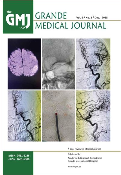
Method: Biopsy of aneurysmal dome and the superficial temporal artery (STA) were done in cases however biopsy of STA taken from those who have undergone surgical procedure like trauma and other cranial pathology. All specimens were obtained during craniotomy. Parameters in vessels biopsy were seen as per histopathological parameters that involves Eccentric fibrointimal thickening, Luminal narrowing, Myxoid degeneration, disruption of internal elastic lamina, medial fibrosis, loss of smooth muscle in media, loss of elastic fibres.
Result: The study was conducted in 27 patients to find out the histopathological parameters of vessels as mentioned above in relation to Connective Tissue Disorder. This was compared with 20 patients with non-aneurysmal pathology. Changes observed in the superficial temporal artery and the dome of the aneurysm in cases of intracranial aneurysms indicate weak connective tissue characteristics.
Conclusion: The histologic changes seen in dome of aneurysm and STA in cases of intra cranial aneurysm a weak connective tissue. In patients with intracranial aneurysms, these changes resemble those seen in CTD. We found notable differences between the two groups in terms of histopathological findings. eccentric thickening of the fibrous inner layer, myxoid degeneration, reduced muscle in the middle layer of the vessel wall, and loss of elastic fibers compared to the control group.