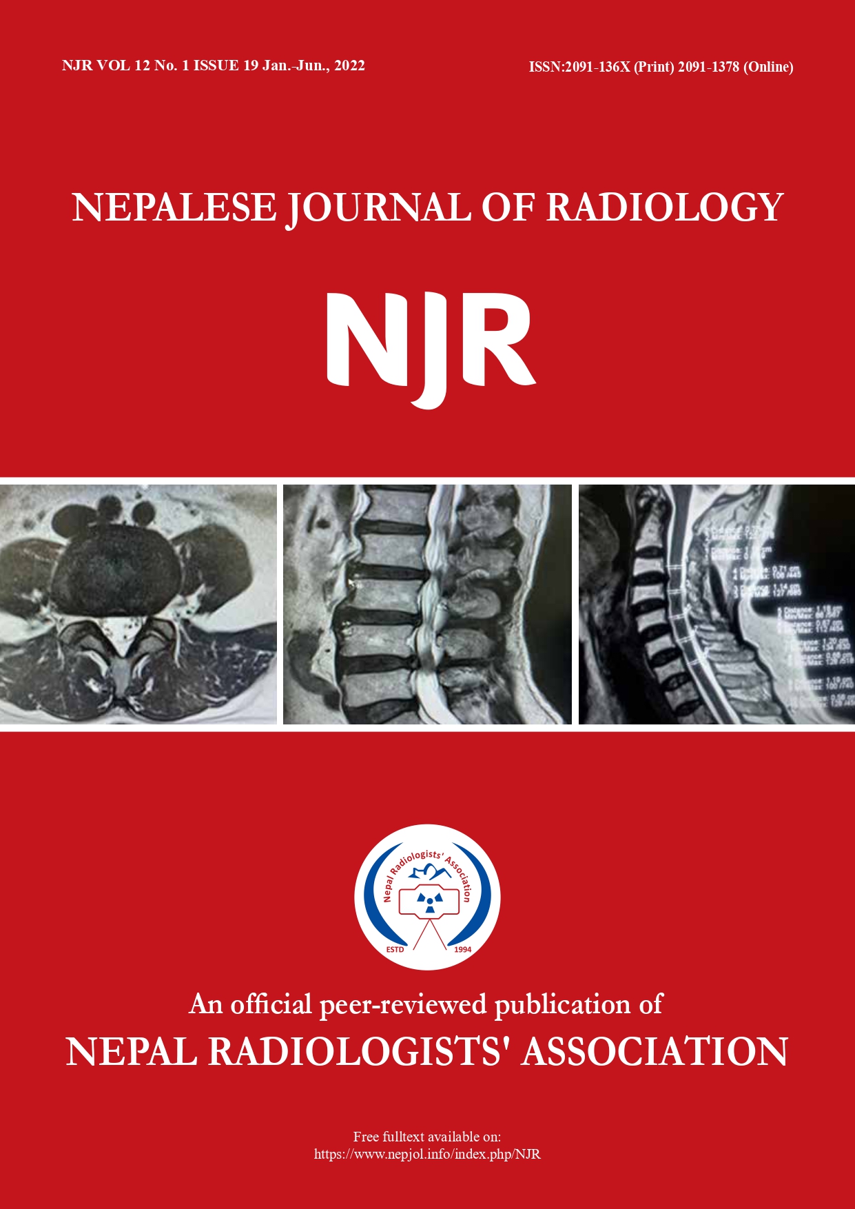
Measurement of Space Available for Cord of Cervical Spine in MRI
DOI:
https://doi.org/10.3126/njr.v12i1.46795Keywords:
Magnetic Resonance Imaging, Spinal Canal, Spinal StenosisAbstract
Introduction: Cervical spinal stenosis has been established as a predisposing factor of cervical myelopathy and is associated with cord injury. Space available for the cord (SAC) can be used as an indicator of spinal stenosis.
Methods: This study was performed on patients referred for MRI examinations of the cervical spine for various clinical indications to a tertiary centre in Nepal. Data were collected for a period of four months from January to April after IRB approval. Convenience sampling was employed and a total of 72 examinations were included. Data were obtained from the 1.5T Magnetom Amira Siemens MRI scanner. Sagittal diameters of the spinal canal and spinal cord were traced and measured from C3 to the C7 vertebra. The space available for the spinal cord (SAC) was calculated by subtracting the sagittal cord diameter from the corresponding sagittal canal diameter.
Results: The average space available for cord was 4.48mm±1.04mm at C3, 4.44mm±1.03mm at C4, 4.63mm±1.01mm at C5, 5.11mm±1.07mm at C6, 5.87mm±1.14mm at C7 vertebral level. The SAC value was not significant according to gender and age (p>0.05).
Conclusions: The smallest SAC value was detected at the C4 vertebral level with a mean value of 4.44mm and the greatest value was at C7 vertebral level with a mean value of 5.87mm. There was no significant gender difference in SAC values.
Downloads
Downloads
Published
How to Cite
Issue
Section
License
Copyright (c) 2022 Nepalese Journal of Radiology

This work is licensed under a Creative Commons Attribution 4.0 International License.
This license enables reusers to distribute, remix, adapt, and build upon the material in any medium or format, so long as attribution is given to the creator. The license allows for commercial use.


