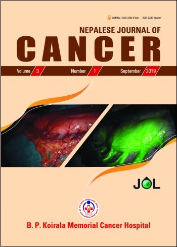
Fluoroscence Angiography for Assessment of Gastric Conduit: Initial Experience from a Tertiary Care Center
DOI:
https://doi.org/10.3126/njc.v3i1.25909Keywords:
Indocyanine green, angiography, fluorescence, esophagectomy, anastomotic leakAbstract
Introduction: Anastomotic leak (AL) after surgery for esophageal cancer remains a main cause of postoperative morbidity and mortality. Poor tissue perfusion at the site of anastomosis is one of the major factors for leak. We aimed to review the results of Indocyanine Green dye (ICG) with a goal to decrease the leak rate.
Methods: Patients with cancer of esophagus and gastroesophageal junction were subjected to either upfront surgery or preoperative chemoradiation/ chemotherapy followed by surgery. Stomach was used for reconstruction. Intravenous injection of 5 mg – 10 mg ICG was given and vascular perfusion was assessed with infrared light of laparoscopic telescope. Gastroesophageal anastomosis was made at the site of adequate ICG perfusion. These patients (ICG group) was compared to the other group of patients in whom ICG was not used (Non-ICG group).
Results: 28 and 396 patients belonged to ICG and Non-ICG group, respectively. 61% in ICG group and 32% in Non-ICG group had preoperative treatment (p <.001). AL was observed in 7% and 16% in ICG and Non-ICG group, respectively (p = 0.2). Healing time of leak was 15 days in ICG group and 32 days in Non-ICG group (p = .03). One patient required revision of anastomotic site based on ICG finding. There was no adverse reaction related to ICG injection.
Conclusion: Fluoroscence angiography using ICG is a safe method for evaluation of vascular perfusion of gastric conduit. Though the leak rate was not statistically different in the two groups, ICG group required lesser time for complete resolution of AL, which might indicate lesser severity of anastomotic disruption.
Downloads
Downloads
Published
How to Cite
Issue
Section
License
This license lets others distribute, remix, tweak, and build upon your work, even commercially, as long as NJC and the authors are acknowledged.
Submission of the manuscript means that the authors agree to assign exclusive copyright to NJC. The aim of NJC is to increase the visibility and ease of use of open access scientific and scholarly articles thereby promoting their increased usage and impact.


